Thursday, December 12, 2013

Methods of Inheritance

For most of my students, this will be a review of stuff you did in Freshman/Sophomore Biology. It is vital background which you will need to build on.  So, if you DON'T know this, you need to learn it.  If it is EASY for you, feel free to skip it.

Try these problems:
1. In mice, the allele for brown fur is dominant over white fur.  A pure breeding brown mouse is mated to a pure breeding white mouse.  What are the expected genotype and phenotype ratios?
2. A mouse from the offspring in problem 1 is mated to a white mouse.  What are the expected phenotype and genotype ratios?
3. Two mice from the offspring of the cross in problem 1 are mated.  What are the expected phenotype and genotype ratios?

[answers are at the end of this post.

If these three problems were difficult for you to answer, you should watch the first 5 minutes of this video from Mr. Anderson at Bozeman Science.  Otherwise, you can continue on to the next problem.


In some cases, two alleles mix to create a phenotype.  These cases are called incomplete dominance and codominance.  In incomplete dominance, the phenotype produced is intermediate.  In codominance, the phenotype produced is a mix of the two.  In reality, this is probably an unimportant distinction, but you can still find it in textbooks and on standardized tests.  Here's a sample problem:

4. Blood types A and B display codominance.  An individual homozygous for blood type A and an individual homozygous for blood type B have kids.  What are the odds that they will have a child with blood type A?  B?  AB?
5. Two individuals with blood type AB mate.  What are the odds that their children will have blood type A? B? AB?

If you had difficulty with problems 4 and 5, begin the video above at about 4:30 and watch the portion about snapdragons.

Some genes are located on the sex chromosomes.  Genes located on the X chromosome are particularly interesting since male humans only get one copy of the X chromosome (termed hemizygous) where as females get two copies.

6. The gene for red-green color vision is located on the X chromosome.  R = normal vision, r = red/green colorblind.  A man with normal vision marries a woman with normal vision whose father was colorblind.  What are the odds of having a color blind boy?  a color blind girl?

If you had difficulty with problem 6, start watching the video at 5:46.

You can get more practice with this type of problem here:
Arizona Monohybrid Crosses
It is vital that you have a good solid foundation with monohybrid crosses.

The Bozeman video goes on to describe how to do dihybrid crosses.  This is great and you should watch it if you aren't familiar with them.  I will teach you a way to do these that is much simpler than writing out 16 square crosses.

We will begin our investigation into inheritance with dihybrid crosses.


Taste genetics

Everyone knows that the tongue allows you to taste.  But how does it work?  It all comes down to ligand-receptor binding.  The diagram below (from learn genetics) shows the connection between the anatomy of the tongue and the cellular location of receptors.

The receptor we are studying in class is the TAS2R38 receptor which binds to the compound phenylthiocarbamide (PTC).  Binding sends signals to the brain which are interpreted as bitter.

What happens if you can't make a working copy of the PTC receptor?

The PTC receptor is a membrane-bound protein made by a ribosome reading an mRNA.
The mRNA is transcribed from DNA found in the nucleus (and then processed).
Every cell in your body has the same DNA in its nucleus.  The PTC gene is expressed (turned on) in tongue cells.

Here is the DNA sequence for the PTC gene:

        1 cctttctgca ctgggtggca accaggtctt tagattagcc aactagagaa gagaagtaga
       61 atagccaatt agagaagtga catcatgttg actctaactc gcatccgcac tgtgtcctat
      121 gaagtcagga gtacatttct gttcatttca gtcctggagt ttgcagtggg gtttctgacc
      181 aatgccttcg ttttcttggt gaatttttgg gatgtagtga agaggcaggc actgagcaac
      241 agtgattgtg tgctgctgtg tctcagcatc agccggcttt tcctgcatgg actgctgttc
      301 ctgagtgcta tccagcttac ccacttccag aagttgagtg aaccactgaa ccacagctac
      361 caagccatca tcatgctatg gatgattgca aaccaagcca acctctggct tgctgcctgc
      421 ctcagcctgc tttactgctc caagctcatc cgtttctctc acaccttcct gatctgcttg
      481 gcaagctggg tctccaggaa gatctcccag atgctcctgg gtattattct ttgctcctgc
      541 atctgcactg tcctctgtgt ttggtgcttt tttagcagac ctcacttcac agtcacaact
      601 gtgctattca tgaataacaa tacaaggctc aactggcaga ttaaagatct caatttattt
      661 tattcctttc tcttctgcta tctgtggtct gtgcctcctt tcctattgtt tctggtttct
      721 tctgggatgc tgactgtctc cctgggaagg cacatgagga caatgaaggt ctataccaga
      781 aactctcgtg accccagcct ggaggcccac attaaagccc tcaagtctct tgtctccttt
      841 ttctgcttct ttgtgatatc atcctgtgtt gccttcatct ctgtgcccct actgattctg
      901 tggcgcgaca aaataggggt gatggtttgt gttgggataa tggcagcttg tccctctggg
      961 catgcagcca tcctgatctc aggcaatgcc aagttgagga gagctgtgat gaccattctg
     1021 ctctgggctc agagcagcct gaaggtaaga gccgaccaca aggcagattc ccggacactg
     1081 tgctgagaat ggacatgaaa tgagctcttc attaatacgc ctgtgagtct tcataaatat
     1141 gcc


This DNA sequence is transcribed into an mRNA.  The mRNA is read to create a protein.  Here is the amino acid sequence of the protein that is a functional receptor.  Note that each letter corresponds to one amino acid.

MLTLTRIRTVSYEVRSTFLFISVLEFAVGFLTNAFVFLVNFWDV
VKRQALSNSDCVLLCLSISRLFLHGLLFLSAIQLTHFQKLSEPLNHSYQAIIMLWMIA
NQANLWLAACLSLLYCSKLIRFSHTFLICLASWVSRKISQMLLGIILCSCICTVLCVW
CFFSRPHFTVTTVLFMNNNTRLNWQIKDLNLFYSFLFCYLWSVPPFLLFLVSSGMLTV
SLGRHMRTMKVYTRNSRDPSLEAHIKALKSLVSFFCFFVISSCVAFISVPLLILWRDK
IGVMVCVGIMAACPSGHAAILISGNAKLRRAVMTILLWAQSSLKVRADHKADSRTLC

Below are five mutations of a single nucleotides that produce non-functional PTC receptor proteins.  I have highlighted these nucleotides in yellow in the DNA sequence above.


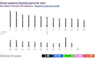
In your nucleus, you have 46 chromosomes in 23 pairs.  One of each pair comes from your mother, the other from your father.  The image below shows the 23 different chromosomes.  The TAS2R38 gene is located on chromosome 7.


Let's imagine that you inherited a copy of functional TAS2R38 from mom as well as one from dad.  We would call you homozygous for the taster allele.  Would you be able to taste PTC?

Let's imagine that you inherited a mutated copy of TAS2R38 from both your mother and your father.  We would say you were homozygous for the nontaster allele.  Would you be able to taste PTC?

Now, let's imagine you were heterozygous.  You got one functional copy from one parent, but a nonfunctional copy from the other.  Would you be able to taste PTC?

You probably already know about dominant and recessive alleles.  Dominant alleles are said to "mask" the presence of recessive alleles.  Hopefully, you answered that heterozygotes can taste PTC (they have the taster phenotype).  That would make the taster allele dominant.

So, the dominant allele is due to a functional copy of a protein, while the recessive allele is a nonfunctional copy.  This is usually the case for genes that show traditional Mendelian dominant/recessive relationships.  

Here is a good video from Howard Hughes Medical Institute on PTC:


Wednesday, December 11, 2013

Structure of genes

We've discussed the structure of genes in class. This activity should help reinforce these concepts and introduce you to some powerful tools in bioinformatics. Identify a protein you are interested in. Search for your protein on Wikipedia.  I choose Wikipedia because it is easy to access and had a lot of information in a format that is presented simply.  We will use it as a jumping off point to the National Center for Biological Information (NCBI) -- which is a reliable source of information.  Because Wikipedia is most complete for human proteins, I encourage you to choose a human protein.

I'm using hemoglobin for my example.  Search Wikipedia for your protein (Hemoglobin). Notice that hemoglobin has 3 subunits. I am going to choose HBA1 for my study. I click on HBA1.


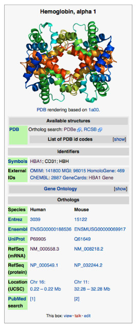
At right is part of the page for Hemoglobin subunit alpha 1.  For the purposes of this exercise, you want to compare mRNA sequences, so click on RefSeq(mRNA)


Scroll down the the bottom of the page that opens and you will find the DNA sequence corresponding to the mRNA as shown below for hemoglobin subunit a.  

This is the mRNA sequence from a eukaryote.  Which of the following does it contain?
  1. Promoter
  2. Start codon
  3. Stop codon
  4. Introns
  5. Exons


ORIGIN      
        1 actcttctgg tccccacaga ctcagagaga acccaccatg gtgctgtctc ctgccgacaa
       61 gaccaacgtc aaggccgcct ggggtaaggt cggcgcgcac gctggcgagt atggtgcgga
      121 ggccctggag aggatgttcc tgtccttccc caccaccaag acctacttcc cgcacttcga
      181 cctgagccac ggctctgccc aggttaaggg ccacggcaag aaggtggccg acgcgctgac
      241 caacgccgtg gcgcacgtgg acgacatgcc caacgcgctg tccgccctga gcgacctgca
      301 cgcgcacaag cttcgggtgg acccggtcaa cttcaagctc ctaagccact gcctgctggt
      361 gaccctggcc gcccacctcc ccgccgagtt cacccctgcg gtgcacgcct ccctggacaa
      421 gttcctggct tctgtgagca ccgtgctgac ctccaaatac cgttaagctg gagcctcggt
      481 ggccatgctt cttgcccctt gggcctcccc ccagcccctc ctccccttcc tgcacccgta
      541 cccccgtggt ctttgaataa agtctgagtg ggcggc
//

Select your sequence and copy it to your clipboard.

Go to the ORF finder at NCBI 

Paste your sequence into the query box and hit OrfFind (the button is oddly placed above the data entry box).  You will get something that looks like this:


My translated sequence is in the +2 frame and is 429 bases long.
Answer these questions for your sequence:
  1. What is shown in blue/green?
  2. How long is the translated portion?
  3. Are there any untranslated portions?
  4. Which frame is translated?

Next, take your sequence and go to nucleotide blast (blastn) at NCBI.
Copy your sequence into the query box.  Select "human genomic + transcript in the database block.
Click blast.

When the results are shown, select "human genome view" from the other views option.

This will take you to a screen which will show you which chromosome your gene is on.


My protein, hemoglobin a is on chromosome 16.


What chromosome is your gene on?


If you click on the chromosome, you will go to the map viewer.  Find a region that shows high identity with a red box in the gene seq map (3rd line) and click on the blue text.  Select "Sequence Viewer".
Selecting sequence viewer will bring up something that looks like this:
Dark green boxes with arrows are exons, light green boxes are untranslated regions of the mRNA and non-boxed green lines are introns.  I can see that Hemoglobin a has two introns and 3 exons.  I can also see the 5' and 3' UTR's.  If I were to scroll to the left on this sequence, I should be able to find the promoter sequence.
  1. How many exons is the gene composed of?
  2. How many introns does it contain?
  3. Can you find your 5'UTR and your 3'UTR?
  4. Where would you expect to find your start and stop codons?


Friday, November 22, 2013

How to Keep Your Online Notebook

Here is a screencast showing you how to create your online lab notebook for AP Biology:


Tuesday, November 19, 2013

Establishing DNA as the Genetic Material

Once upon a time, we didn't know that DNA is the genetic material.  There were some really cool experiments that people did to figure out what was going on.  I have summarized them in this screencast:


Here is a good video on the history of the discovery of the structure of DNA:


Here is a good video on the process of replication:


Sunday, November 17, 2013

Graphing data over time and finding rates of reactions

In our Photosynthesis and Enzymes labs, we looked at change in transmittance or absorbance over time to measure the progress of a reaction.  This screencast will tell you what to do with your data.

Friday, November 8, 2013

Breathing and Gas Exchange.

Below are parts 1 and 2 of a screencast I did on Breathing and Gas Exchange

Tuesday, September 24, 2013

How Plants Control their "Breathing"

We've talked about how water moves through a plant and introduced the idea that gas exchange happens through the stomata.  Most people look at plants and see non-moving things; but plants actually have many moving parts.  One of these is the stomata.  Stomata are holes in the plant leaf that are surrounded by guard cells.  Plants regulate the size of this opening by osmotically swelling or shrinking the guard cells.

What gases pass through stomata?

  • Carbon dioxide (in during the day, out during night)
  • Oxygen (out during the day, in during the night)
  • Water (out at all times)
Plants typically close their stomata at night--not all the way closed of course since they still need to get rid of carbon dioxide from respiration.  The receptor turns out to respond to blue wavelengths of light.  When you shine blue light onto the plant, the receptor triggers a signal cascade that results in the guard cells becoming turgid and the stomata open.


The mechanism for opening relies on a proton pump setting up a concentration gradient of H+ across the cell membrane.

 It uses this proton gradient to drive a K+ into the cell.  This lowers water potential in the guard cell causing water to enter.  The cells swell, opening the stomata.  Closing them seems to be a result of simply releasing the K+ by facilitated diffusion.  The exact signaling pathways that control stomata are an area of active research.  In any case, here is a fun demonstration of how stomata open and close.


Monday, September 23, 2013

From the Soil to the Sky Part II - Transpirational Pull

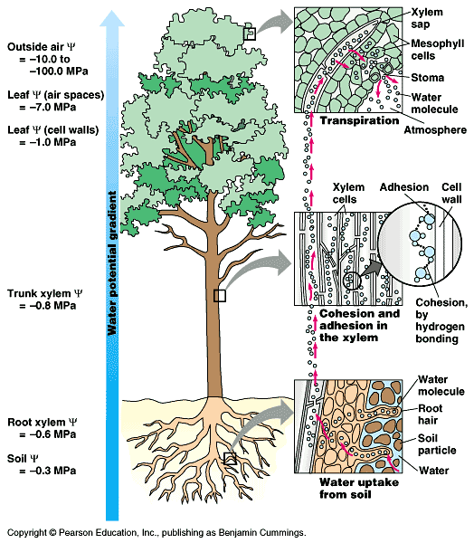
In the previous post, we saw that root pressure is only enough to account for water rising 7 meters.  In a tree that is 112 m tall, we still have 105 meters to go.  Using the football field analogy, we aren't even out of our own end zone using root pressure.  Transpirational pull accounts for the rest.

Water flows downhill.  It flows from where there is a lot of water to where there isn't a lot of water.  We have seen that water will flow into a cell as long as the solute concentration of the cell is higher than the external environment.  We call this cell hypertonic to its' hypotonic environment.  We say that the water potential of the cell is lower (or more negative) than the environment.  To put it in terms of water potential, water flows from high to low water potential.

Since we are talking about pulling water, we need to start where the water will be pulled from.


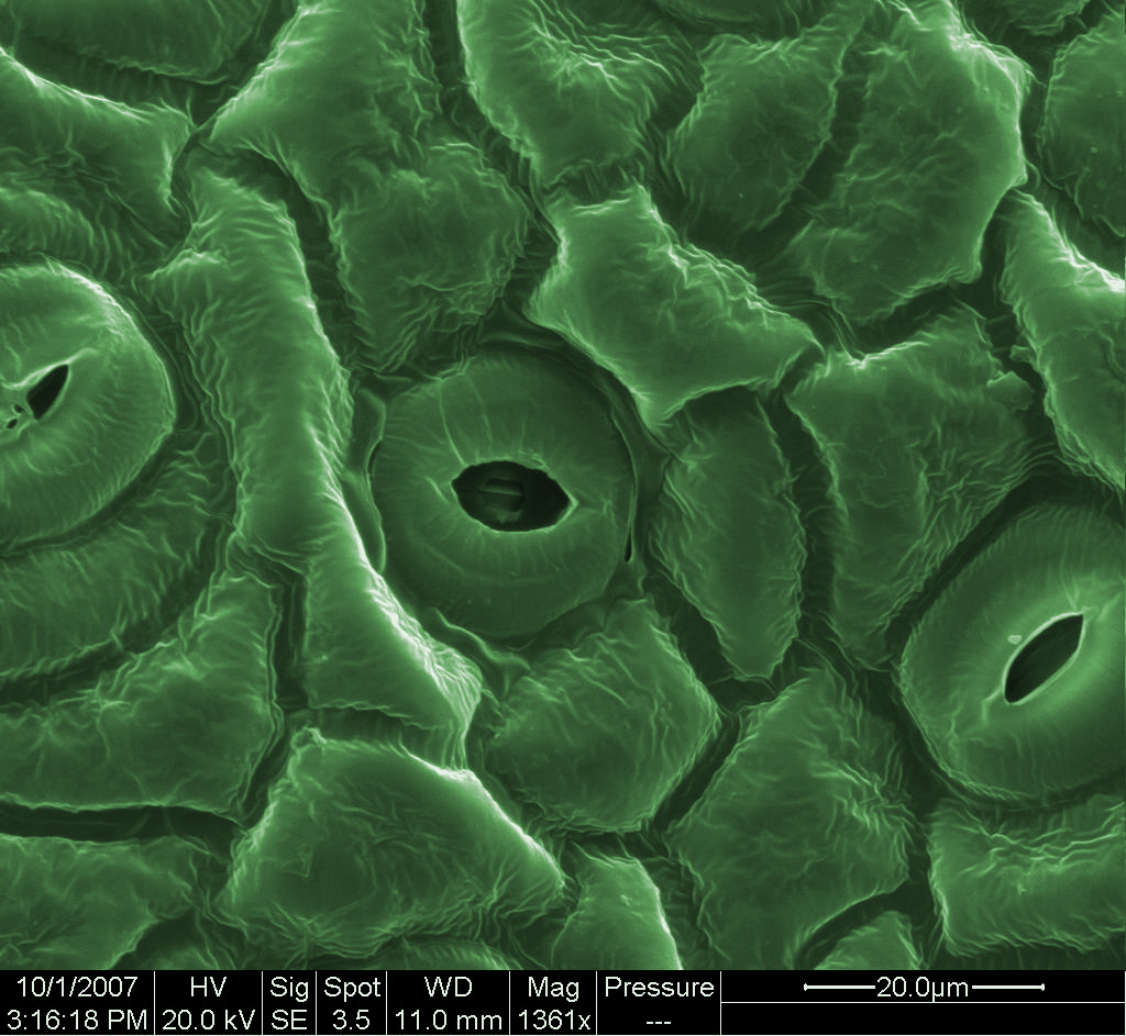
Leaves have openings in them called stomata which are created by two cells called guard cells (shown above).  Plants can adjust these openings so that they are wide open, partly open or even tightly closed (more on the mechanism later).  Most stomata are on the undersides of leaves.  Gases including carbon dioxide, oxygen and water vapor are exchanged through the stomata.


Leaf SEM


Leaf cross section (light microscope)


In the cross-section of a leaf above, you can see that the stomatal opening allows gases into the spongy mesophyll, which is an air-filled cavity in the leaf surrounding plant cells.  Each of these cells is exposed to the air, and is also connected to other cells which are connected to veins.  Veins contain xylem cells (pictured below) which carry water.


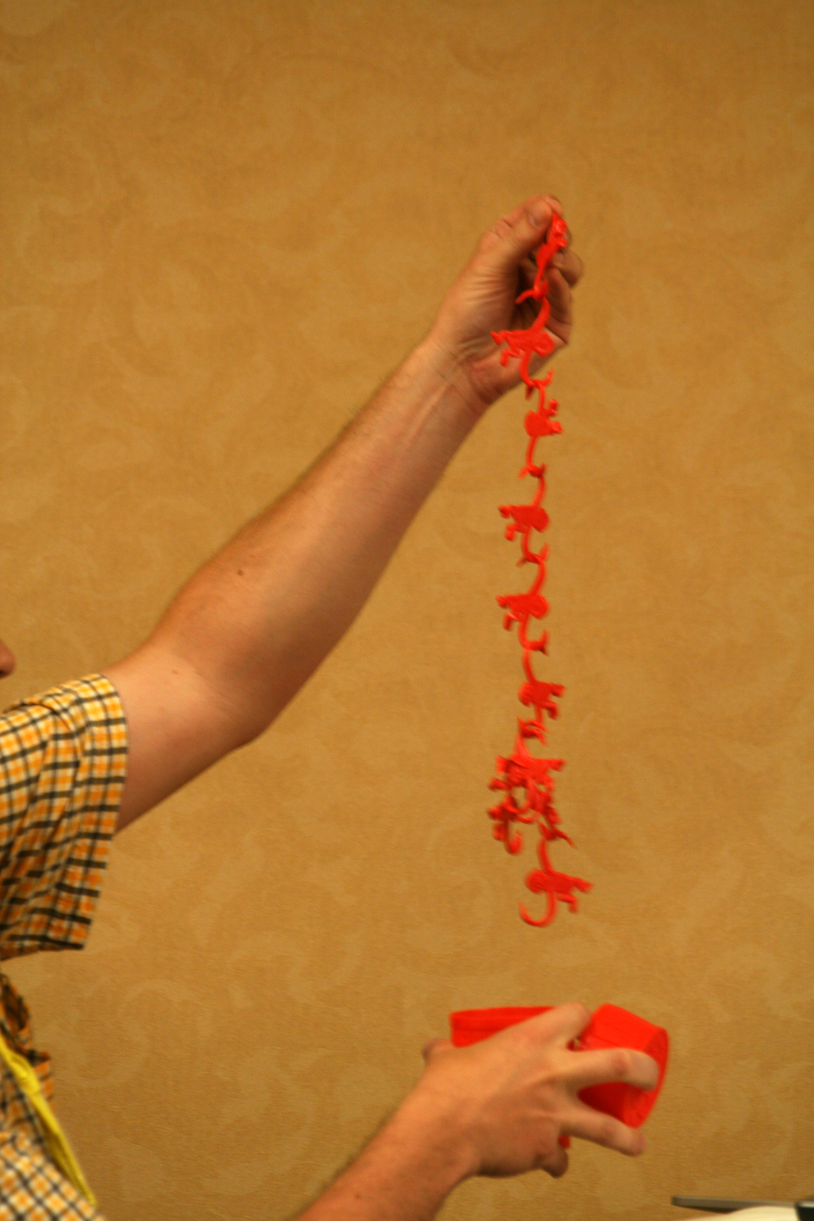
Wood is xylem.  Water travels in a continuous fashion from the spongy mesophyll all the way down the plant stem in the xylem.  Each water molecule hydrogen bonds to the ones around (and below) them--this is cohesion.  When one molecule evaporates into the air space in the leaf, other molecules are pulled up to take its place.  Because they are all connected, each water molecule pulls on the one below it, much like in the game barrel of monkeys.

Cohesion of water is like a barrel of monkeys.

Water flows into the air from the plant because there is less water there--the water potential is lower.  Depending on the humidity, air may be between -10 and -100 MPa.  As we move into the stomatal cavity, humidity goes up and water potential also becomes closer to zero (higher).  As long as the water potential is progressively higher as we go down the tree to the root, water will continue to flow from the roots up the stem to the leaf and out the stomata.


Remember that life is in a dynamic state of disequilibrium.  As the soil drys out, the plant will do things to conserve water and to maintain its internal water potential lower than the soil.  At some point this will be impossible and the plant.

What factors do you suppose affect the rate of transpiration (the loss of water from a leaf surface)?  How could you measure transpiration?  How would you design an experiment to test the effect of one of these factors?


Friday, September 20, 2013

From the Soil to the Sky - How Water Moves


We’ve talked about water potential on the level of a single cell, and that would probably suffice if all we cared about were algae.  But land plants are much more complex, and interesting.  David Attenborough explains the problem in this video.


So how do you get water from the soil up to the leaf surface where you will use the water for photosynthesis and to transport necessary nutrients?   There really are only two ways to do this—you either push or pull.

It turns out that plants do both, but that one is much stronger than the other.  We’ll examine root pressure first and then transpirational pull.

To understand root pressure, you have to know something about transport across cell membranes.  Here’s a good video:


Of course, if you want to understand how a root works, you need to know something about its’ anatomy.


Look at the diagram first and see what you can deduce.

Things you should notice:

1.     Plant cells are connected by holes in their cell walls through which the plasma membranes are continuous.  These holes are called plasmodesmata.  You will notice that, once water passes a plasma membrane, it can travel unobstructed through the cells into the interior of the plant.
2.     Water does not have to pass a membrane at the root at a root hair.  It can enter at any point before the central ring.  This central ring of cells is called the endodermis.  They secrete a hydrophobic substance (a wax) that prevents water from moving between the cells of the endodermis.
3.     This makes the root cells one huge, continuous folded membrane in contact with the external environment (extremely high surface area to volume ratio).

Here’s a picture of a real root


The Y-shaped things are root hairs.  They are covered with fungi.  These fungi are called mycorrhizae and are mutualistic symbionts (90% of plant species have mycorrhizal partners) .  They help the plant with water and nutrient uptake in exchange for sugars.

Compare the diameter of a root hair (which is a single plant cell) with the diameter of one of the cottony fungal strands.  What does this do to the surface area:volume ratio?

----
You should now realize that the root in combination with a mycorrhizal fungus is basically one huge membrane.  Now imagine that you put H+ pumps all along this membrane.  Along with these H+ pumps, you put coporters that allow the H+ ions back in, but bring along with them some nutrients like NO32-, PO43-.  What now happens to the osmotic conditions inside the endodermis?  Is it hypertonic or hypotonic to the soil?  Is the water potential higher or lower?

The osmotic conditions generated are enough to cause guttation on plants in wet soil in the morning. 


Root pressure is only enough to pump water up about 7 meters under the best conditions. 


The tallest tree in the world is 112 meters tall  (a football field including both end zones (122 yards).  Transpirational pull makes up the difference.  That will be the topic of my next post.

Tuesday, September 17, 2013

Atomsmith Demonstration

Atomsmith is a really cool program, but doesn't seem to work for everyone. A very computer-savvy student could probably contact the authors and help to de-bug it for multiple platforms. It really is a program that should be used in every science class.

Since many of my students have had problems with it, and my non-student readers don't have access to the program, I recorded a screencast of some of the important parts of the summer assignment I wrote. Here it is:

Monday, June 24, 2013

Pingry Community Research Journal

I'm pleased to share with you Pingry's newest publication--a journal dedicated to publishing experimental research conducted by our students.


PCR is a student-written and student-edited.  This is the first edition.  My AP Biology students will want to read through it since they will be expected to complete a research project and write a paper for publication in this journal.  I'm hopeful that others will want to read some of the articles simply because they are interesting.  Click on the image above to go to a .pdf version of the journal.

Wednesday, June 19, 2013

Zombie Cockroaches, Killer Fungi and Evolving Mousetraps


I was thinking about the mousetrap analogy that refutes the idea of Irreducible Complexity.  If you aren't familiar with it, here it is:


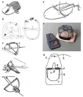
One thing that always bothered me about Miller's argument is that the mousetrap didn't, in fact, evolve.  So, could it have evolved?  There are many examples of traps in the natural world.  I thought I'd collect a few for you.

Killer Fungi - Cordyceps are a group of fungi that infect insects and take over their minds.


Zombie Cockroach - More mind control.  Here's a species of wasp that injects a neurochemical into the cockroach to allow the wasp to guide it into a grave.  After laying eggs on the still-live cockroach, the wasp seals the tomb.


The Ant Lion - an animal that makes a trap for insects.    


Fungi that make lassos


Venus flytrap - I think you'd have to call this an evolved trap.


And the grand finale - an actual mousetrap that evolved:


The mousetrap we are all familiar with was the product of people's imagination and there have been many variations, most of which never made money (went extinct).  The simplest trap really is a small stick holding something heavy up.  You put the bait on the stick.  When the mouse eats the bait, the roof falls.  Here's an interesting paper discussing it called Exploring Mouse Trap History