This is an assignment for my freshman biology class, but I think it would be appropriate for anyone who is interested in learning about proteins. Here is the assignment:
Snow day work:
1. Choose a protein. Any protein will do, though some are easier than others.
2. Go to the class spreadsheet on google drive and type the name of the protein you chose next to your name.
https://docs.google.com/spreadsheet/ccc?key=0At0IcHkYPpqBdFF1U19Gc21DenNWZXpvUXJtcVhlV1E&usp=sharing
3. In a document file, answer the following:
a. Name of the protein
b. How many subunits is it made of?
c. Give the primary structure (amino acid sequence) of one of the polypeptide subunits (you may use the single letter abbreviation for amino acids.
d. Take a screenshot of your protein showing a secondary structure (alpha helix or beta pleated sheet for example.
e. Does your protein make any disulfide bridges between cystines? If so, take a screenshot.
f. Are there any nonpolar regions? Where and what do they do?
g. Does your protein have an active site? A channel? Show something interesting about your protein.
I will post an example of what I'm looking for on my blog (http://biologuy.blogspot.com/). You must at least attempt this assignment. If you get stuck, post questions here. If there are no questions here and you tell me that you got stuck, we have a problem. Spend no more than 45 minutes on this exercise.
Rubisco is my favorite protein since it is responsible for making plants from thin air (carbon dioxide levels are about 390 parts per million right now (
current CO2). How do you find a protein to choose? The Protein Data Bank has a wonderful section called the
Molecule of the Month. Browse through these until you find one you're interested in. Wikipedia also has good information.
Rubisco at the
PDB MOM
Rubisco at
wikipedia
a.Rubisco is an abbreviation for
Ribulose-1,5-bisphosphate carboxylase/oxygenase.
b.In terrestrial (land) plants, Rubisco has an L8S8 structure, which means that there are 8 large subunits and 8 small subunits.
c. To find the primary structure, or the amino acid sequence, you will most likely have to go to the pdb file. Here's how. In the molecule of the month article, you can find a link to the coordinate file. In the image below, the entry 1rcx links to the information we want.
When you click on 1rcx, it takes you to a page that looks like this
If you click on the tab that says "sequence" you will see something that looks like this:
The primary sequence begins with a methionine (M) and ends with a tyrosine (Y). You could simply type the primary sequence from this screen; but there is a fasta way -- click on FASTA.
Here's mine:
>1RCX:C|PDBID|CHAIN|SEQUENCE
MQVWPILNLKKYETLSYLPPLTTDQLARQVDYLLNNKWVPCLEFETDHGFVYREHHNSPGYYDGRYWTMWKLPMFGCTDPAQVLNELEECKKEYPNAFIRIIGFDSNREVQCISFIAYKPAGY
Now, why am I making you do this? So that you will understand what a primary sequence is. Each letter represents a different amino acid. Primary structure is the amino acid sequence.
d. Take a screenshot showing a secondary structure. You should be able to see from the image above that Rubisco has 2 alpha helices (the wavy red lines) and some beta sheets (the yellow arrows). I want you to show me a 3-d view of these though. If you click on one of the alpha helices or open the 3-d view tab, it will open a program called jmol. You may have to install or update java to get it to work--I did--DON'T install the lame ASK toolbar though.
Here's a 3-d view of an alpha helix in Rubisco:
e. Does Rubisco have any disulfide bridges? The easiest way to find disulfide bridges is to right-click (or command click) and color>disulfide bonds. If you can't see them, there may not be any or you may not be looking right. To be sure, right click and select>protein>by residue name>CYS. This will select all cystines (the amino acid responsible for disulfide bridges). Then select>display selected only. This will remove everything from your view except for cystines that make disulfide bridges. Rubisco appears to have no disulfide bridges.
f. Nonpolar regions - you can find these by select>protein>nonpolar residues and then select>display selected only.
g. Does your protein have an active site? A channel? Show something interesting about your protein. Answering this question will depend largely on your protein. For Rubisco, the active site can be found by select>hetero>ligand and then select>display selected only. The image below shows 8 ligands (so 8 active sites).
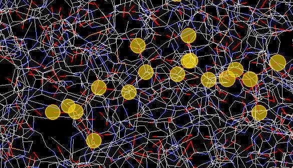
To show the area around one of these ligands (the active site), I chose select>all then style>scheme>wireframe. Then I chose select>hetero>ligand and then select>selection halos and took this picture:
The halos are surrounding individual atoms in the ligand.
Here's another shot with the halos off and the ligand in ball-and-stick with the protein in wireframe:
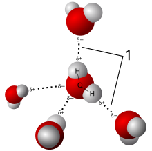
In this image, you can see the 5-carbon sugar (carbons in gray) with two phosphate groups (P = yellow, O = Red).