Friday, February 20, 2015

Zombie Cockroaches, Killer Fungi and Evolving Mousetraps Updated


I was thinking about the mousetrap analogy that refutes the idea of Irreducible Complexity.  If you aren't familiar with it, here it is:


One thing that always bothered me about Miller's argument is that the mousetrap he's using in his analogy didn't, in fact, evolve.  So, could it have evolved?  There are many examples of traps in the natural world.  I thought I'd collect a few for you.

Killer Fungi - Cordyceps are a group of fungi that infect insects and take over their minds. 


Zombie Cockroach - More mind control.  Here's a species of wasp that injects a neurochemical into the cockroach to allow the wasp to guide it into a grave.  After laying eggs on the still-live cockroach, the wasp seals the tomb.


The Ant Lion - an animal that makes a trap for insects.    


Fungi that make lassos to capture little worms called nematodes


Venus flytrap - I think you'd have to call this an evolved trap.


And the grand finale - an actual mousetrap that evolved:


The mousetrap we are all familiar with was the product of people's imagination and there have been many variations, most of which never made money (went extinct).  The simplest trap really is a small stick holding something heavy up (or you could use the bait to hold the rock up).  You put the bait on the stick.  When the mouse eats the bait, the roof falls.  Here's an interesting paper discussing it called Exploring Mouse Trap History


Tuesday, December 9, 2014

Inheritance Essentials

For most of my students, this will be a review of stuff you did in Freshman/Sophomore Biology. It is vital background which you will need to build on.  So, if you DON'T know this, you need to learn it.  If it is EASY for you, feel free to skip it.

Try these problems:
1. In mice, the allele for brown fur is dominant over white fur.  A pure breeding brown mouse is mated to a pure breeding white mouse.  What are the expected genotype and phenotype ratios?
2. A mouse from the offspring in problem 1 is mated to a white mouse.  What are the expected phenotype and genotype ratios?
3. Two mice from the offspring of the cross in problem 1 are mated.  What are the expected phenotype and genotype ratios?

[answers are at the end of this post.]

If these three problems were difficult for you to answer, watch the following video.  Otherwise, you can continue on to the next problem.


In some cases, two alleles mix to create a phenotype.  These cases are called incomplete dominance and codominance.  In incomplete dominance, the phenotype produced is intermediate.  In codominance, the phenotype produced is a mix of the two.  In reality, this is probably an unimportant distinction, but you can still find it in textbooks and on standardized tests.  Here's a sample problem:

4. Blood types A and B display codominance.  An individual homozygous for blood type A and an individual homozygous for blood type B have kids.  What are the odds that they will have a child with blood type A?  B?  AB?
5. Two individuals with blood type AB mate.  What are the odds that their children will have blood type A? B? AB?

If you had difficulty with problems 4 and 5, watch this video:


Some genes are located on the sex chromosomes.  Genes located on the X chromosome are particularly interesting since male humans only get one copy of the X chromosome (termed hemizygous) where as females get two copies.

6. The gene for red-green color vision is located on the X chromosome.  R = normal vision, r = red/green colorblind.  A man with normal vision marries a woman with normal vision whose father was colorblind.  What are the odds of having a color blind boy?  a color blind girl?

If you had difficulty with problem 6, watch this video:


You can get more practice with this type of problem here:
Arizona Monohybrid Crosses
It is vital that you have a good solid foundation with monohybrid crosses.

We will begin our investigation into inheritance with dihybrid crosses.


Tuesday, July 1, 2014

Summer Assignment 2014-15

AP Biology is over-enrolled this year at Pingry.  Because we do not have enough teachers to cover 3 sections, all Juniors have been wait-listed.  If you are a Senior who is not fully committed to the class, consider dropping to make space for a dedicated Junior.  The class will move rapidly.  I will expect you to come to class in September with the following assignments completed.

Before you begin any work, complete the preinstruction test.

I will email your results to you.  Once you have your results, complete the preinstruction test analysis form.

Complete the following assignments


Chemistry of Biology - Using Avogadro to Investigate Organic Chemicals


Essay:  Chemistry of Large Biological Molecules


Atomsmith tutorial

  If you can get Atomsmith to install on your computer, do this tutorial.

  Most people will have difficulty getting this to install.  If this is your situation, view this video to answer the questions in the tutorial.


Expect to have a test on this information shortly after the fall semester begins (within the first two weeks).  


Monday, February 3, 2014

Exploring Protein Structure

This is an assignment for my freshman biology class, but I think it would be appropriate for anyone who is interested in learning about proteins.  Here is the assignment:

Snow day work:
1. Choose a protein.  Any protein will do, though some are easier than others.
2. Go to the class spreadsheet on google drive and type the name of the protein you chose next to your name.
https://docs.google.com/spreadsheet/ccc?key=0At0IcHkYPpqBdFF1U19Gc21DenNWZXpvUXJtcVhlV1E&usp=sharing
3. In a document file, answer the following:
a. Name of the protein
b. How many subunits is it made of?
c. Give the primary structure (amino acid sequence) of one of the polypeptide subunits (you may use the single letter abbreviation for amino acids.
d. Take a screenshot of your protein showing a secondary structure (alpha helix or beta pleated sheet for example.
e. Does your protein make any disulfide bridges between cystines?  If so, take a screenshot.
f. Are there any nonpolar regions?  Where and what do they do?
g. Does your protein have an active site?  A channel?  Show something interesting about your protein.

I will post an example of what I'm looking for on my blog (http://biologuy.blogspot.com/).  You must at least attempt this assignment.  If you get stuck, post questions here.  If there are no questions here and you tell me that you got stuck, we have a problem.  Spend no more than 45 minutes on this exercise.

Rubisco is my favorite protein since it is responsible for making plants from thin air (carbon dioxide levels are about 390 parts per million right now (current CO2).  How do you find a protein to choose?  The Protein Data Bank has a wonderful section called the Molecule of the Month.  Browse through these until you find one you're interested in.  Wikipedia also has good information.
Rubisco at the PDB MOM
Rubisco at wikipedia

a.Rubisco is an abbreviation for Ribulose-1,5-bisphosphate carboxylase/oxygenase.
b.In terrestrial (land) plants, Rubisco has an L8S8 structure, which means that there are 8 large subunits and 8 small subunits.
c. To find the primary structure, or the amino acid sequence, you will most likely have to go to the pdb file.  Here's how.  In the molecule of the month article, you can find a link to the coordinate file.  In the image below, the entry 1rcx links to the information we want.


When you click on 1rcx, it takes you to a page that looks like this

If you click on the tab that says "sequence" you will see something that looks like this:

The primary sequence begins with a methionine (M) and ends with a tyrosine (Y).  You could simply type the primary sequence from this screen; but there is a fasta way -- click on FASTA.

Here's mine:
>1RCX:C|PDBID|CHAIN|SEQUENCE
MQVWPILNLKKYETLSYLPPLTTDQLARQVDYLLNNKWVPCLEFETDHGFVYREHHNSPGYYDGRYWTMWKLPMFGCTDPAQVLNELEECKKEYPNAFIRIIGFDSNREVQCISFIAYKPAGY

Now, why am I making you do this?  So that you will understand what a primary sequence is.  Each letter represents a different amino acid.  Primary structure is the amino acid sequence.

d. Take a screenshot showing a secondary structure.  You should be able to see from the image above that Rubisco has 2 alpha helices (the wavy red lines) and some beta sheets (the yellow arrows).  I want you to show me a 3-d view of these though.  If you click on one of the alpha helices or open the 3-d view tab, it will open a program called jmol.  You may have to install or update java to get it to work--I did--DON'T install the lame ASK toolbar though.

Here's a 3-d view of an alpha helix in Rubisco:

e. Does Rubisco have any disulfide bridges?  The easiest way to find disulfide bridges is to right-click (or command click) and color>disulfide bonds.  If you can't see them, there may not be any or you may not be looking right.  To be sure, right click and select>protein>by residue name>CYS.  This will select all cystines (the amino acid responsible for disulfide bridges).  Then select>display selected only.  This will remove everything from your view except for cystines that make disulfide bridges.  Rubisco appears to have no disulfide bridges.

f. Nonpolar regions - you can find these by select>protein>nonpolar residues and then select>display selected only.

g. Does your protein have an active site?  A channel? Show something interesting about your protein.  Answering this question will depend largely on your protein.  For Rubisco, the active site can be found by select>hetero>ligand and then select>display selected only.  The image below shows 8 ligands (so 8 active sites).


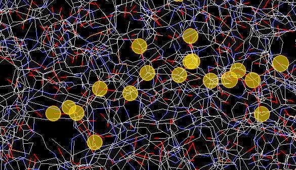
To show the area around one of these ligands (the active site), I chose select>all then style>scheme>wireframe.  Then I chose select>hetero>ligand and then select>selection halos and took this picture:


The halos are surrounding individual atoms in the ligand.
Here's another shot with the halos off and the ligand in ball-and-stick with the protein in wireframe:


In this image, you can see the 5-carbon sugar (carbons in gray) with two phosphate groups (P = yellow, O = Red).

Thursday, December 12, 2013

Methods of Inheritance

For most of my students, this will be a review of stuff you did in Freshman/Sophomore Biology. It is vital background which you will need to build on.  So, if you DON'T know this, you need to learn it.  If it is EASY for you, feel free to skip it.

Try these problems:
1. In mice, the allele for brown fur is dominant over white fur.  A pure breeding brown mouse is mated to a pure breeding white mouse.  What are the expected genotype and phenotype ratios?
2. A mouse from the offspring in problem 1 is mated to a white mouse.  What are the expected phenotype and genotype ratios?
3. Two mice from the offspring of the cross in problem 1 are mated.  What are the expected phenotype and genotype ratios?

[answers are at the end of this post.

If these three problems were difficult for you to answer, you should watch the first 5 minutes of this video from Mr. Anderson at Bozeman Science.  Otherwise, you can continue on to the next problem.


In some cases, two alleles mix to create a phenotype.  These cases are called incomplete dominance and codominance.  In incomplete dominance, the phenotype produced is intermediate.  In codominance, the phenotype produced is a mix of the two.  In reality, this is probably an unimportant distinction, but you can still find it in textbooks and on standardized tests.  Here's a sample problem:

4. Blood types A and B display codominance.  An individual homozygous for blood type A and an individual homozygous for blood type B have kids.  What are the odds that they will have a child with blood type A?  B?  AB?
5. Two individuals with blood type AB mate.  What are the odds that their children will have blood type A? B? AB?

If you had difficulty with problems 4 and 5, begin the video above at about 4:30 and watch the portion about snapdragons.

Some genes are located on the sex chromosomes.  Genes located on the X chromosome are particularly interesting since male humans only get one copy of the X chromosome (termed hemizygous) where as females get two copies.

6. The gene for red-green color vision is located on the X chromosome.  R = normal vision, r = red/green colorblind.  A man with normal vision marries a woman with normal vision whose father was colorblind.  What are the odds of having a color blind boy?  a color blind girl?

If you had difficulty with problem 6, start watching the video at 5:46.

You can get more practice with this type of problem here:
Arizona Monohybrid Crosses
It is vital that you have a good solid foundation with monohybrid crosses.

The Bozeman video goes on to describe how to do dihybrid crosses.  This is great and you should watch it if you aren't familiar with them.  I will teach you a way to do these that is much simpler than writing out 16 square crosses.

We will begin our investigation into inheritance with dihybrid crosses.


Taste genetics

Everyone knows that the tongue allows you to taste.  But how does it work?  It all comes down to ligand-receptor binding.  The diagram below (from learn genetics) shows the connection between the anatomy of the tongue and the cellular location of receptors.

The receptor we are studying in class is the TAS2R38 receptor which binds to the compound phenylthiocarbamide (PTC).  Binding sends signals to the brain which are interpreted as bitter.

What happens if you can't make a working copy of the PTC receptor?

The PTC receptor is a membrane-bound protein made by a ribosome reading an mRNA.
The mRNA is transcribed from DNA found in the nucleus (and then processed).
Every cell in your body has the same DNA in its nucleus.  The PTC gene is expressed (turned on) in tongue cells.

Here is the DNA sequence for the PTC gene:

        1 cctttctgca ctgggtggca accaggtctt tagattagcc aactagagaa gagaagtaga
       61 atagccaatt agagaagtga catcatgttg actctaactc gcatccgcac tgtgtcctat
      121 gaagtcagga gtacatttct gttcatttca gtcctggagt ttgcagtggg gtttctgacc
      181 aatgccttcg ttttcttggt gaatttttgg gatgtagtga agaggcaggc actgagcaac
      241 agtgattgtg tgctgctgtg tctcagcatc agccggcttt tcctgcatgg actgctgttc
      301 ctgagtgcta tccagcttac ccacttccag aagttgagtg aaccactgaa ccacagctac
      361 caagccatca tcatgctatg gatgattgca aaccaagcca acctctggct tgctgcctgc
      421 ctcagcctgc tttactgctc caagctcatc cgtttctctc acaccttcct gatctgcttg
      481 gcaagctggg tctccaggaa gatctcccag atgctcctgg gtattattct ttgctcctgc
      541 atctgcactg tcctctgtgt ttggtgcttt tttagcagac ctcacttcac agtcacaact
      601 gtgctattca tgaataacaa tacaaggctc aactggcaga ttaaagatct caatttattt
      661 tattcctttc tcttctgcta tctgtggtct gtgcctcctt tcctattgtt tctggtttct
      721 tctgggatgc tgactgtctc cctgggaagg cacatgagga caatgaaggt ctataccaga
      781 aactctcgtg accccagcct ggaggcccac attaaagccc tcaagtctct tgtctccttt
      841 ttctgcttct ttgtgatatc atcctgtgtt gccttcatct ctgtgcccct actgattctg
      901 tggcgcgaca aaataggggt gatggtttgt gttgggataa tggcagcttg tccctctggg
      961 catgcagcca tcctgatctc aggcaatgcc aagttgagga gagctgtgat gaccattctg
     1021 ctctgggctc agagcagcct gaaggtaaga gccgaccaca aggcagattc ccggacactg
     1081 tgctgagaat ggacatgaaa tgagctcttc attaatacgc ctgtgagtct tcataaatat
     1141 gcc


This DNA sequence is transcribed into an mRNA.  The mRNA is read to create a protein.  Here is the amino acid sequence of the protein that is a functional receptor.  Note that each letter corresponds to one amino acid.

MLTLTRIRTVSYEVRSTFLFISVLEFAVGFLTNAFVFLVNFWDV
VKRQALSNSDCVLLCLSISRLFLHGLLFLSAIQLTHFQKLSEPLNHSYQAIIMLWMIA
NQANLWLAACLSLLYCSKLIRFSHTFLICLASWVSRKISQMLLGIILCSCICTVLCVW
CFFSRPHFTVTTVLFMNNNTRLNWQIKDLNLFYSFLFCYLWSVPPFLLFLVSSGMLTV
SLGRHMRTMKVYTRNSRDPSLEAHIKALKSLVSFFCFFVISSCVAFISVPLLILWRDK
IGVMVCVGIMAACPSGHAAILISGNAKLRRAVMTILLWAQSSLKVRADHKADSRTLC

Below are five mutations of a single nucleotides that produce non-functional PTC receptor proteins.  I have highlighted these nucleotides in yellow in the DNA sequence above.


In your nucleus, you have 46 chromosomes in 23 pairs.  One of each pair comes from your mother, the other from your father.  The image below shows the 23 different chromosomes.  The TAS2R38 gene is located on chromosome 7.


Let's imagine that you inherited a copy of functional TAS2R38 from mom as well as one from dad.  We would call you homozygous for the taster allele.  Would you be able to taste PTC?

Let's imagine that you inherited a mutated copy of TAS2R38 from both your mother and your father.  We would say you were homozygous for the nontaster allele.  Would you be able to taste PTC?

Now, let's imagine you were heterozygous.  You got one functional copy from one parent, but a nonfunctional copy from the other.  Would you be able to taste PTC?

You probably already know about dominant and recessive alleles.  Dominant alleles are said to "mask" the presence of recessive alleles.  Hopefully, you answered that heterozygotes can taste PTC (they have the taster phenotype).  That would make the taster allele dominant.

So, the dominant allele is due to a functional copy of a protein, while the recessive allele is a nonfunctional copy.  This is usually the case for genes that show traditional Mendelian dominant/recessive relationships.  

Here is a good video from Howard Hughes Medical Institute on PTC:


Wednesday, December 11, 2013

Structure of genes

We've discussed the structure of genes in class. This activity should help reinforce these concepts and introduce you to some powerful tools in bioinformatics. Identify a protein you are interested in. Search for your protein on Wikipedia.  I choose Wikipedia because it is easy to access and had a lot of information in a format that is presented simply.  We will use it as a jumping off point to the National Center for Biological Information (NCBI) -- which is a reliable source of information.  Because Wikipedia is most complete for human proteins, I encourage you to choose a human protein.

I'm using hemoglobin for my example.  Search Wikipedia for your protein (Hemoglobin). Notice that hemoglobin has 3 subunits. I am going to choose HBA1 for my study. I click on HBA1.


At right is part of the page for Hemoglobin subunit alpha 1.  For the purposes of this exercise, you want to compare mRNA sequences, so click on RefSeq(mRNA)


Scroll down the the bottom of the page that opens and you will find the DNA sequence corresponding to the mRNA as shown below for hemoglobin subunit a.  

This is the mRNA sequence from a eukaryote.  Which of the following does it contain?
  1. Promoter
  2. Start codon
  3. Stop codon
  4. Introns
  5. Exons


ORIGIN      
        1 actcttctgg tccccacaga ctcagagaga acccaccatg gtgctgtctc ctgccgacaa
       61 gaccaacgtc aaggccgcct ggggtaaggt cggcgcgcac gctggcgagt atggtgcgga
      121 ggccctggag aggatgttcc tgtccttccc caccaccaag acctacttcc cgcacttcga
      181 cctgagccac ggctctgccc aggttaaggg ccacggcaag aaggtggccg acgcgctgac
      241 caacgccgtg gcgcacgtgg acgacatgcc caacgcgctg tccgccctga gcgacctgca
      301 cgcgcacaag cttcgggtgg acccggtcaa cttcaagctc ctaagccact gcctgctggt
      361 gaccctggcc gcccacctcc ccgccgagtt cacccctgcg gtgcacgcct ccctggacaa
      421 gttcctggct tctgtgagca ccgtgctgac ctccaaatac cgttaagctg gagcctcggt
      481 ggccatgctt cttgcccctt gggcctcccc ccagcccctc ctccccttcc tgcacccgta
      541 cccccgtggt ctttgaataa agtctgagtg ggcggc
//

Select your sequence and copy it to your clipboard.

Go to the ORF finder at NCBI 

Paste your sequence into the query box and hit OrfFind (the button is oddly placed above the data entry box).  You will get something that looks like this:


My translated sequence is in the +2 frame and is 429 bases long.
Answer these questions for your sequence:
  1. What is shown in blue/green?
  2. How long is the translated portion?
  3. Are there any untranslated portions?
  4. Which frame is translated?

Next, take your sequence and go to nucleotide blast (blastn) at NCBI.
Copy your sequence into the query box.  Select "human genomic + transcript in the database block.
Click blast.

When the results are shown, select "human genome view" from the other views option.

This will take you to a screen which will show you which chromosome your gene is on.


My protein, hemoglobin a is on chromosome 16.


What chromosome is your gene on?


If you click on the chromosome, you will go to the map viewer.  Find a region that shows high identity with a red box in the gene seq map (3rd line) and click on the blue text.  Select "Sequence Viewer".
Selecting sequence viewer will bring up something that looks like this:
Dark green boxes with arrows are exons, light green boxes are untranslated regions of the mRNA and non-boxed green lines are introns.  I can see that Hemoglobin a has two introns and 3 exons.  I can also see the 5' and 3' UTR's.  If I were to scroll to the left on this sequence, I should be able to find the promoter sequence.
  1. How many exons is the gene composed of?
  2. How many introns does it contain?
  3. Can you find your 5'UTR and your 3'UTR?
  4. Where would you expect to find your start and stop codons?